Showing posts with label Case Studies. Show all posts

Monday, 29 June 2009

Case 3 (b)

This post follows the previous one, where we were introduced to a 71-year-old man with symptoms and signs that were suspicious for colorectal cancer.

We left off with the question: what does the chest X-ray show?

The principle finding on the man's chest X-ray is multiple, well-defined, round opacities in each lung field. The most likely diagnosis here is multiple lung metastases. Other, less likely possibilities include infectious causes (multiple abscesses, septic emboli, fungal infiltrates), inflammatory diseases (rheumatoid nodules, nodules from Wegener's granulomatosis or sarcoid), or (rarely) pulmonary arteriovenous malformations.


Of course, we already had our suspicions as to where to go looking for the primary tumour. Nonetheless:

Question 4: In the context of multiple pulmonary metastases, what are the commonest primary sites?

The four most likely culprits are probably: bronchial, breast, gastrointestinal tract, and renal cancers.


The patient's full (complete) blood count came back as follows:
  • White Cell Count: 9.0 x 10⁹/ L [Normal: 4-11]
  • Haemoglobin: 7.7 g/dL [Normal: 13.5 - 16.5]
  • MCV: 69 fL [Normal: 79-95]
  • Platelets: 414 x 109/L [Normal 150-450]

Question 5: Comment of the full blood count. What is the most likely cause of this abnormality?

The patient has a microcytic anaemia (with a normal white cell count and platelet count). The most common cause of this is iron deficiency anaemia. This ties in nicely with a possible diagnosis of colorectal carcinoma, since these cancers often bleed (usually microscopically) over many months, causing the loss of iron from the body.

The patient underwent a colonoscopy which showed what appeared to be a broad-based tumour at the splenic flexure. The subsequent histology report indicated it to be a colorectal adenocarcinoma. Prognosis is, sadly, poor.

Sunday, 28 June 2009

Case 3 - Spot Diagnosis

A 71 year-old man presented to casualty with the non-specific symptoms of weight loss (from about 80 kg to 70 kg over four months), anorexia, constipation and fatigue. These symptoms had been gradually increasing in intensity over several months.

Question 1: What is anorexia?

Students often confuse the symptom of anorexia with the medical condition "anorexia nervosa". Anorexia is simply an inappropriately depressed appetite. (This definition distinguishes the condition from that "full" feeling or satiety that you have after wolfing down a massive hamburger.)

Question 2: What is particularly worrying about this man's symptom constellation?

An elderly man with a chronic change in bowel habit should reflexly prompt you to consider the possibility of colon cancer, even if another diagnosis is more likely. In this man's case, there are additional "red flags" that make this diagnosis a distinct possibility: weight loss and anorexia.

On examination the man was noted to be wasted, and had conjunctival pallor. He had no significant lymphadenopathy or organomegally, and his cardiovascular, respiratory and neurological examinations were normal apart from a tachycardia of 102 bpm. His abdomen was slightly distended, but there was no evidence of peritonism and bowel sounds were normal. A rectal exam was normal.

In addition to bloods, both chest and abdominal X-rays were ordered. Below is (something very similar to) his chest X-ray:


Question 3: What does the chest X-ray show?

We'll answer that tomorrow.

Thursday, 21 May 2009

Case 2: the answer

Thanks to those who put forward their prospective answers, either via email or in the comments section.  This was the X-ray of a 65-year-old smoker.

The X-ray shows three notable things:
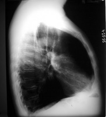
  • The anterior-posterior diameter of the chest looks enlarged and "barrel-shaped".
  • There is an increase in the amount of air 'trapped' anterior to the cardiac shadow.
  • The diaphragms are flattened.
Together these point to generalised hyperinflation.  On the AP (or PA) view you would presumably see an increase in the number of ribs visible.

There are several causes of generalised hyperinflation, but the most likely of these, in a 65-year-old smoker, is chronic obstructive pulmonary disease (COPD).  Other possibilities for generalised hyperinflation are:
  • Asthma (when symptomatic)
  • Cystic fibrosis
  • α1-antitrypsin deficiency
  • endobronchial sarcoidosis

Tuesday, 19 May 2009

Case 2: Spot diagnosis

Comment on the following lateral chest X-ray, and state the most likely diagnosis in this 65-year-old smoker.


I'll post the answer tomorrow.

Wednesday, 29 April 2009

Case 1: an update

Remember the young woman with severe B12 deficiency? I saw her again yesterday; she's doing really well. Her haemoglobin is up to 9.6 g/dl (up, on B12 supplements alone, from about 4), the rest of her full blood count has normalised, and she tells me she's eating like a horse! (No doubt this is the increased caloric demand from her body's cells suddenly being able to divide properly again, plus the absence of the nausea that had contributed to her weight loss.)

Her anti-parietal cell antibodies have come back negative, and the anti-intrinsic factor ones are unaccountably still not back yet, which doesn't exactly help us in terms of confirming pernicious anaemia (although they aren't always positive in this condition either). We've decided to do another gastroscopy with a view to getting a biopsy of the stomach, which should settle the matter. We've also done a brief screen for concomitant disorders of other endocrine glands (e.g. thyroid, adrenal cortex), which can be simultaneously dysfunctional in certain cases of pernicious anaemia.


UPDATE 02/05/09

Her anti-intrinsic factor antibody assay has come back positive, which helps point towards our original diagnosis of pernicious anaemia.

Sunday, 19 April 2009

Case 1(b)

[This follows on from the first part of the case]

The patient's full blood count was as follows (normal ranges are in brackets):
  • White Cell Count: 1.2 x 10⁹/l  (4-11)
  • Haemoglobin: 2.4 g/dl  (11.5-16.5)
  • MCV [mean corpuscular volume]: 112 fl (80-95)
  • Platelets: 23 x 109/l (150-450)

Question 4: How does the blood count reconcile the patient's haematemesis with her normal gastroscopy findings?

The patient has a severe pancytopenia, including a severe thrombocytopenia.  The latter causes a bleeding tendency, which may occassionally present as an upper GIT bleed in the absence of significant ulcers or erosions.


Question 5: Are there any clues as to the aetiology of the pancytopenia from the blood count?

Yes - the anaemia is macrocytic (the MCV is markedly raised).  This raises the possibility that we are dealing with a megaloblastic anaemia, which is caused by a deficiency or folate and/or vitamin B12.  A severe enough deficiency of one of these vitamins can cause a pancytopenia.


Sure enough, the peripheral blood smear showed oval (as opposed to round) macrocytes and hypersegmented neutrophils.  Together with the raised MCV, this is virtually diagnostic of a megaloblastic anaemia, and hence possibly the pancytopenia by extension.  As further blood results poured in, the folate level was found to be normal, but the B12 level was only about a third of the lower limits of normality.  We had our presumptive diagnosis: pancytopenia secondary to a severe vitamin B12 deficiency.  


Question 6: What are the causes of B12 deficiency?

We've answered that, in passing, here.


The rest of the patient's presenting pathologies then fell into place:
  • Her weight loss turned out to be from a long-standing combination of gastrointestinal symptoms including nausea and diarrhoea.  There was probably an element of malabsorption too.  These aren't infrequent symptoms in megalobastic anaemias, since like the haematological cells, the mucosa's turnover rate is extremely rapid and thus very sensitive to a nutrients.
  • Careful examination revealed a glossitis (in which the normal tongue papillae are absent), which is an extension of the GIT involvement in general.
  • The ejection systolic murmur, by the way, was due to the patient's high cardiac output state, which was secondary to her severe anaemia in turn.  The increased rate of ejection of an increased volume of blood causes turbulence around the aortic outlet, even though the actual valve is anatomically normal.  Why do you get a high cardiac output state in anaemia?  See here
Fortunately, the patient hadn't developed any neurological symptoms (such as paraesthesias, numbness or defects of proprioception), since these are often permanent.


Question 7:  Why does a vitamin B12 deficiency affect the blood and gastrointestinal mucosal cells most?

A deficiency of vitamin B12 affects all cells that divide, since it leads to an inability to synthesize adequate amounts of DNA.  (Cells that divide each have to take a copy of the body's DNA with them, and so must replicate it before dividing.) It turns out that vitamin B12 is a vital cofactor in the synthesis of thymidine (T) from uridine (U).  Therefore, without it cells can't make DNA (thymidine is one of the nucleosides, remember?), and therefore can't divide at the appropriate rate.  The commonest symptoms (haematological and gastrointestinal) simply reflect the fact that these cells divide the quickest, and so any slowing of cellular division is likely to be noticed there first.


Question 8:  Why does vitamin B12 deficiency cause neurological dysfunction?  Those cells either don't divide quickly or at all!

Indeed, neurological dysfunction in B12 deficiency is by another mechanism (or two).  In addition to being a cofactor in normal thymidine synthesis, B12 is also a cofactor in the synthesis of an amino acid called methionine.  Although the exact pathogenetic chain is yet to be worked out, it seems as if the deficiency of methionine causes demyelination (loss of the normal myelin sheaths around the nerves).  Furthermore, succinyl-CoA production is also slowed in B12 deficiency.  The fact that this compound is involved in fatty acid synthesis may also partly account for the demyelination.


The patient was supplemented with B12 by means of a series of intramuscular injections.  She responded beautifully, with a rapid response in all three cell lines.  Within days, both her platelets and her white cells were at normal levels, and her anaemia was well on the road to oblivion.

Currently, she is being worked up as to the cause of her B12 deficiency.  Could it be pernicious anaemia?  None of the other likely causes of B12 deficiency seemed to fit.  The gastroscopy findings didn't mention gastric atrophy, but I doubt if this was specifically looked for at 2 o'clock in the morning when we thought we were dealing with a severe upper gastrointestinal bleed alone.  Two antibodies associated with pernicious anaemia might shed some light on the matter - anti-parietal cell antibody, and anti-intrinsic factor antibody.  These results are currently pending.


Question 9:  How does pernicious anaemia cause B12 deficiency?

'Pernicious anaemia' is the result of an autoimmune attack on the stomach's parietal cells.  As a result of the loss of parietal cell mass, stomach acid production falls to virtually nothing (achlorhydria) and insufficient intrinsic factor is produced.  Both of these facts cause a decrease in vitamin B12 absorption.  A low pH is necessary to release B12 from any ingested food, and intrinsic factor's job is to bind B12, thereby preventing its degradation, and enhancing its absorption in the terminal ileum. 

That about does it for the first case.  Comments on the format, anyone?  Good, bad, indifferent?

Saturday, 18 April 2009

Case 1(a)

Mrs S was a 34 year-old female who presented to casualty with three episodes of haematemesis over the past week.  She thought that she vomited about 50 mls of frank blood each time.  She had no other past medical history, nor did she have a recent history of any NSAID [non-steroidal anti-inflammatory drug] or alcohol usage.  She had never noted any malaena stools and did not suffer from dyspepsia or heartburn.  She wasn't a smoker, was unemployed, and had no significant family history or travel history.

On examination, she was noted to be cachectic, and exhibited severe pallor.  Her blood pressure was 90/48, with a pulse of 105.  The rest of her vitals were normal, as was the rest of her examination, other than a 3/6 ejection systolic murmer heard best over the aortic and pulmonary areas.  The murmur didn't radiate.  


Question 1: What are the commonest causes of haematesis?

The top few causes are:
  • gastric ulcers (25%)
  • duodenal ulcers (25%)
  • erosive gastritis (20%)
  • Mallory-Weis tear (10%), and
  • oesophageal varices (5%).  
As noted from the percentages, there are other causes too, however.


An arterial blood gas analysis was done, and the relevant results are as follows:

  • pH: 7.36
  • pO2 : 80 mmHg (10.6 kPa) [Note: this is normal for the altitude at which it was recorded]
  • pCO2: 29 mmHg (3.86 kPa)
  • Base excess: -7.5
  • Lactate: 4.0
  • Haemoglobin: 2.3

Question 2: Comment on the blood gas analysis.

The results indicate a metabolic acidosis, with respiratory compensation (so that the pH is within normal limits).  The most likely cause of the metabolic acidosis is a lactic acidosis, as the lactate is raised. The patient has an extremely severe anaemia.


The patient was dripped, and Ringer's lactate was infused.  Blood was then ordered, and the patient was sent to the acute medical admissions ward.  [Why not to the surgical acute admissions ward?  I've no idea.  There's always the perennial fight between medicine and surgery, but upper GIT bleeds should be surgical until proven otherwise.]  Here, we transfused three units of packed cells, but were careful to pull bloods before the transfusion, so as to get an accurate reflection of things like iron stores, B12, folate and reticulocytes.


Question three: What is the most likely reason for the lactate to be elevated?

There are many reasons for hyperlactataemia, but anaerobic metabolism is likely to be the one here.  A significant upper GIT bleed would deplete the intravascular blood volume, leading to hypovolaemia.  This would cause inadequate tissue perfusion.  Since blood carries oxygen, a lack of blood to a particular tissue means that the tissue also won't get enough oxygen.  As a result of this, the tissue's cells must perform their metabolic functions as best they can by (the vastly more inefficient) anaerobic metabolism.

However, there is another possibility, given that the haemoglobin is so low.  Oxygen dissolves rather poorly in blood, and so we use haemoglobin to bind to it.  This hugely increases the amound of oxygen carried by the blood.  A very low haemoglobin therefore means that the blood's oxygen content is much reduced.  Therefore, even with adequate tissue perfusion, the body's cells still might not be getting enough oxygen for aerobic metabolism.  As before, they will therefore have to switch to anaerobic metabolism.

A gastroscopy was performed soon thereafter, which was... entirely normal.  No ulcers, no erosions, no blood (old or new) whatsoever, anywhere from the the oesophagus to the proximal duodenum.  This was unexpected, but we soon received the patient's blood results, after which things all made sense.  We'll cover that in the second part of this case.